analytical weighing balance is very important for laboratories that need to be very precise in their measurements of tiny amounts, for example, in clinical, pharmaceutical and research environments. It also helps very well in the accurate preparation of reagents, solutions and samples for analysis. The laboratory staff depends on analytical weighing balance for keeping the same quality, method validation and getting accurate results. Its sensitivity, dependability, and reproducibility make it a necessary tool not only for hospital laboratories but also for research institutes and pharmaceutical quality control processes.
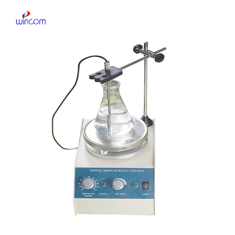
In microbiology labs, analytical weighing balance is utilized for the preparation of culture media and analytical additives. Accurate weighing guarantees that the nutrient composition for microbial growth and testing is consistent. This application helps to produce reliable cultures, conduct antimicrobial studies, and do infection research. By keeping the mass control precise, analytical weighing balance supports reproducibility in microbiological workflows in clinical laboratories.
At the medical institutions that are research-driven, analytical weighing balance will change to facilitate the analytical methods with higher sensitivity that are in the pipe. The future might bring along the possibility of ultra-low mass samples being accurately measured in molecular diagnostics and sophisticated drug research. This turning development will not only enlarge the experimental capacities of hospital-based research labs but also open new fronts in medical innovation through analytics.
Most care routines for analytical weighing balance consist of planned startup and shutdown methods. Giving enough time for the warm-up process to take place guarantees that internal electronics will be operating at stable conditions. Power cuts are not allowed and this way sensitive circuits are protected. The critics of the hospitals that have implemented standard operating procedures operate daily lab analyses with a consistent measure of accuracy and at the same time prolong the life of the analytical weighing balance.
analytical weighing balance is employed in hospital labs for the reliable quality control of reagents, chemicals, and medications. Its exactness provides accurate concentrations for assays, patient treatments, and experimental protocols. The laboratory personnel regularly calibrate analytical weighing balance to rule out mistakes. Its application keeps the standard of hospital laboratories, allows the reproducibility, and builds trust in clinical and research outcomes.
Q: What distinguishes an Analytical Balance from a precision balance? A: The analytical balances have a higher sensitivity and a finer readability for measuring masses of very small amounts. Q: Is an Analytical Balance appropriate for pharmaceutical applications? A: It is widely used for weighing active ingredient and formulation components. Q: Is it mandatory for an Analytical Balance to have a draft shield? A: Draft shields have the function to prevent air disturbances which might affect the weighing results. Q: What are the possible types of materials that can be weighed on an Analytical Balance? A: Weighing of powders, chemicals, and biological samples, as well as reference weights are the most common measurement. Q: Is it possible for several users to work with the same Analytical Balance? A: Yes, but the proper handling procedures and access controls must be strictly adhered to.
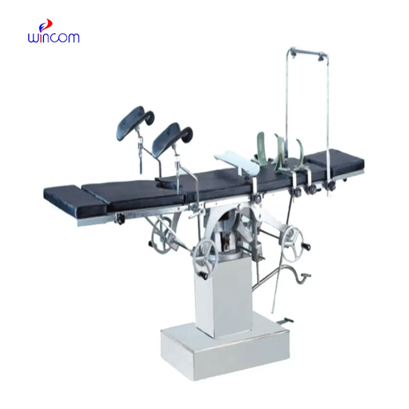
The delivery bed is well-designed and reliable. Our staff finds it simple to operate, and patients feel comfortable using it.
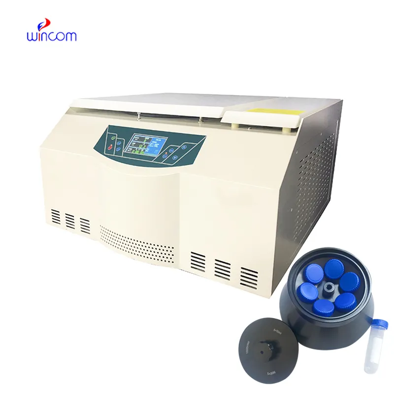
The centrifuge operates quietly and efficiently. It’s compact but surprisingly powerful, making it perfect for daily lab use.
To protect the privacy of our buyers, only public service email domains like Gmail, Yahoo, and MSN will be displayed. Additionally, only a limited portion of the inquiry content will be shown.
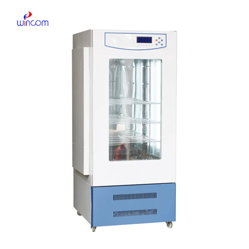
Hello, I’m interested in your water bath for laboratory applications. Can you confirm the temperat...
I’d like to inquire about your x-ray machine models. Could you provide the technical datasheet, wa...
E-mail: [email protected]
Tel: +86-731-84176622
+86-731-84136655
Address: Rm.1507,Xinsancheng Plaza. No.58, Renmin Road(E),Changsha,Hunan,China

 af
af
 es
es
 ar
ar
 tr
tr
 sw
sw
 pt
pt
 th
th
 ur
ur
 bn
bn
 ne
ne
 vi
vi
 km
km
 lo
lo
 de
de
 ru
ru
 fi
fi
 nl
nl
 fa
fa
 fr
fr
 ko
ko