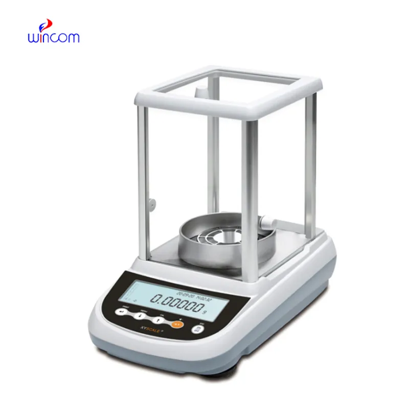
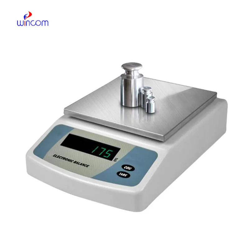
antique analytical balance is, first of all, a crucial instrument in a lab that can provide the accurate measurement of small amounts of things, thus it coalesces into a main character in the laboratory world. That was all hospitals and research labs expect from its precision - the preparation of reagents, patient samples, and medicines. its steady execution guarantees results that can be repeated, helps to ascertain the quality of the analysis, and reinforces the clinical workflows. antique analytical balance is the most important tool for achieving laboratory credibility, experiment precision, and medical and research patient safety in case of of medical and research applications.
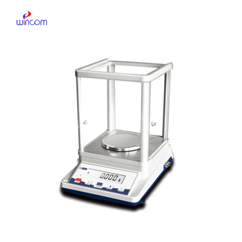
antique analytical balance are used in clinical laboratories for making calibration references that are then checked on the analytics machines. Exact weighing guarantees that the calibration materials keep the same mass values throughout the uses they are subjected to. This application endorses instrument accuracy checks, routine audits in laboratories, and compliance with government regulations. By being a part of trustworthy calibration workflows, antique analytical balance is a major factor and, thus, a contributor to measurement accuracy kept in hospitals' diagnostic devices.
The future of antique analytical balance in medical labs will put more focus on environmental stability. The advanced vibration suppression and temperature control capabilities will support precise operation even in the most crowded hospital areas. This change will make it possible to locate antique analytical balance nearer to the clinical workstations and this, in turn, will result in a reduction of sample transport time. Rather than moving to simpler hospital environments, antique analytical balance will continue to provide quick analytical preparation support and will also maintain high measurement consistency.
The manner in which samples are handled is of utmost importance in the preservation of antique analytical balance. It is necessary for the operators to wear gloves or use instruments to place samples in such a way that contamination and static charges would not be a problem. Regular training of personnel on the proper way to handle instruments reduces physical strain and hence, increases the life of the equipment. Following the right procedures in handling brings about the reliability of the antique analytical balance even in the most demanding hospital laboratories.
To formulate a drug, antique analytical balance is used by the pharmaceutical laboratories to weigh active ingredients and excipients. Precise and reliable measurements not only guarantee the correctness of the dosage but also satisfy the requirements of the regulators. The laboratory personnel use antique analytical balance for quality control, batch verification, and stability testing. Its accuracy aids in the production of medicines that are reliable, thus minimizing errors in the production. Adoption of antique analytical balance into workflow has helped pharmaceutical labs not only to keep their quality standards high but also to make sure that patients are safe by providing the exact analytical measurements.
Q: What distinguishes an Analytical Balance from a precision balance? A: The analytical balances have a higher sensitivity and a finer readability for measuring masses of very small amounts. Q: Is an Analytical Balance appropriate for pharmaceutical applications? A: It is widely used for weighing active ingredient and formulation components. Q: Is it mandatory for an Analytical Balance to have a draft shield? A: Draft shields have the function to prevent air disturbances which might affect the weighing results. Q: What are the possible types of materials that can be weighed on an Analytical Balance? A: Weighing of powders, chemicals, and biological samples, as well as reference weights are the most common measurement. Q: Is it possible for several users to work with the same Analytical Balance? A: Yes, but the proper handling procedures and access controls must be strictly adhered to.
I’ve used several microscopes before, but this one stands out for its sturdy design and smooth magnification control.
The centrifuge operates quietly and efficiently. It’s compact but surprisingly powerful, making it perfect for daily lab use.
To protect the privacy of our buyers, only public service email domains like Gmail, Yahoo, and MSN will be displayed. Additionally, only a limited portion of the inquiry content will be shown.
I’m looking to purchase several microscopes for a research lab. Please let me know the price list ...
Could you please provide more information about your microscope range? I’d like to know the magnif...
E-mail: [email protected]
Tel: +86-731-84176622
+86-731-84136655
Address: Rm.1507,Xinsancheng Plaza. No.58, Renmin Road(E),Changsha,Hunan,China

 af
af
 es
es
 ar
ar
 tr
tr
 sw
sw
 pt
pt
 th
th
 ur
ur
 bn
bn
 ne
ne
 vi
vi
 km
km
 lo
lo
 de
de
 ru
ru
 fi
fi
 nl
nl
 fa
fa
 fr
fr
 ko
ko