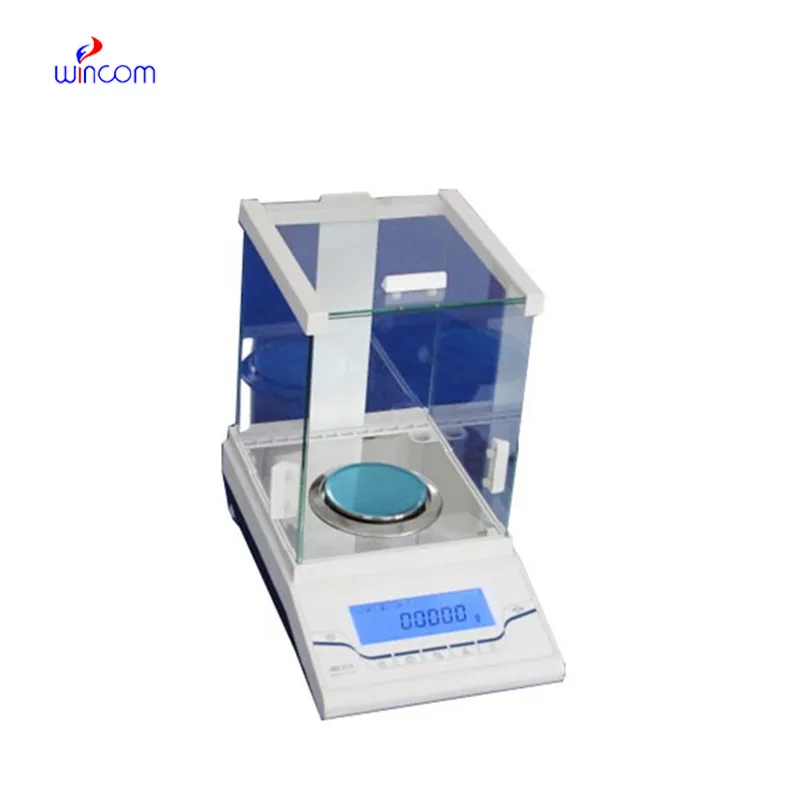
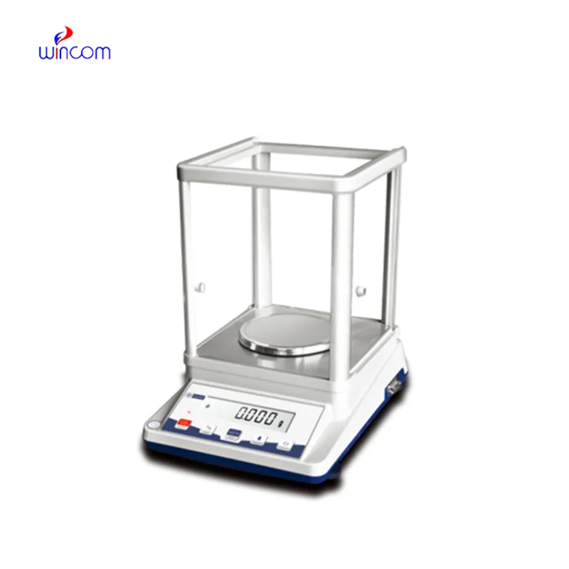
digital analytical balance is very important for laboratories that need to be very precise in their measurements of tiny amounts, for example, in clinical, pharmaceutical and research environments. It also helps very well in the accurate preparation of reagents, solutions and samples for analysis. The laboratory staff depends on digital analytical balance for keeping the same quality, method validation and getting accurate results. Its sensitivity, dependability, and reproducibility make it a necessary tool not only for hospital laboratories but also for research institutes and pharmaceutical quality control processes.
In microbiology labs, digital analytical balance is utilized for the preparation of culture media and analytical additives. Accurate weighing guarantees that the nutrient composition for microbial growth and testing is consistent. This application helps to produce reliable cultures, conduct antimicrobial studies, and do infection research. By keeping the mass control precise, digital analytical balance supports reproducibility in microbiological workflows in clinical laboratories.
The future of digital analytical balance in medical labs will put more focus on environmental stability. The advanced vibration suppression and temperature control capabilities will support precise operation even in the most crowded hospital areas. This change will make it possible to locate digital analytical balance nearer to the clinical workstations and this, in turn, will result in a reduction of sample transport time. Rather than moving to simpler hospital environments, digital analytical balance will continue to provide quick analytical preparation support and will also maintain high measurement consistency.
For digital analytical balance to last long, professionals must do scheduled servicing as per the laboratory guidelines. Internal cleaning and checking of parts will help to slow down and eventually stop the performance degradation. Hospitals or labs that maintain structured service intervals not only enjoy lesser downtimes but also higher accuracy, thus they can carry on with seamless analytical operations.
The precision of digital analytical balance is achieved only in a very controlled environment, which implies regulation of temperature, humidity, and vibration to a minimum level. These parameters are continuously monitored by laboratory technicians to avoid any errors in measurements. digital analytical balance technique provides highly accurate weighing of tiny samples in severe conditions, thus supporting laboratory experiments and hospital-grade analyses of sensitive tests or research that demands careful sample handling.
Q: What is the main purpose of an Analytical Balance? A: Its purpose is mainly to measure very tiny sample masses with the utmost precision in laboratories and hospitals. Q: What is the typical weighing range of an Analytical Balance? A: The weighing range for the majority of analytical balances is from 0 up to some grams with a resolution of micrograms or milligrams. Q: What environmental controls are necessary for an Analytical Balance's operation? A: Airflow, vibration, and temperature changes should not only be avoided but also prevented in the room where the scale is situated. Q: Is an Analytical Balance permitted in a hospital laboratory? A: Yes, it has indeed found widespread usage for the preparation of reagents, calibra¬tion, and drug development applications. Q: What should be the frequency of calibration for an Analytical Balance? A: The calibration interval is subject to the degree of use and the particular laboratory requirements.
The hospital bed is well-designed and very practical. Patients find it comfortable, and nurses appreciate how simple it is to operate.
The microscope delivers incredibly sharp images and precise focusing. It’s perfect for both professional lab work and educational use.
To protect the privacy of our buyers, only public service email domains like Gmail, Yahoo, and MSN will be displayed. Additionally, only a limited portion of the inquiry content will be shown.
We’re looking for a reliable centrifuge for clinical testing. Can you share the technical specific...
Hello, I’m interested in your water bath for laboratory applications. Can you confirm the temperat...
E-mail: [email protected]
Tel: +86-731-84176622
+86-731-84136655
Address: Rm.1507,Xinsancheng Plaza. No.58, Renmin Road(E),Changsha,Hunan,China

 af
af
 es
es
 ar
ar
 tr
tr
 sw
sw
 pt
pt
 th
th
 ur
ur
 bn
bn
 ne
ne
 vi
vi
 km
km
 lo
lo
 de
de
 ru
ru
 fi
fi
 nl
nl
 fa
fa
 fr
fr
 ko
ko