With the cutting-edge imaging processors, the home fetal doppler facilitates real-time, high-resolution images that are paramount in the detection of subtle physiological changes by clinicians. The display is user-friendly for easy parameter modification as well as image marking. The home fetal doppler exhibits the mix of effectiveness, mobility, and reliability for a huge range of diagnostic procedures.

The vast clinical applications of the home fetal doppler technology made it possible for nephrology to monitor kidney function efficiently and detect abnormalities in kidney structure. In the frontiers of endocrinology, the obtained data can reveal even the smallest nodules in the glands. The home fetal doppler is also a surgical device for blood flow patterns and vessel integrity.

The home fetal doppler should integrate with intelligent diagnostic ecosystems and communicate effortlessly with smartphones and electronic records. The synchronized exchange of data in real-time should enable constant patient observation. The next version should focus on improved design, better processing power of artificial intelligence algorithms, and enhanced reconstruction functions.

The daily upkeep of the home fetal doppler involves cleaning, inspection, and proper storage. The removal of the gel residue from the probes should be accomplished as soon as the analysis has been carried out. The cooling vents of the device should always be unblocked. The home fetal doppler needs annual professional servicing in order to remain accurate.
The home fetal doppler uses state-of-the-art ultrasound technology to deliver real-time imaging for diagnostic and monitoring purposes. It aids physicians in assessing organs, blood vessels, and soft tissue with unmatched clarity. The non-surgical device is an important tool for guiding medical procedures and making precise diagnoses. The home fetal doppler combines portability with precision, rendering it extremely useful in routine exams as well as emergency applications.
Q: What is the primary function of an ultrasound scannert? A: Ultrasound scanners are designed to create real-time images of internal organs, tissues, and blood flow using high-frequency sound waves. Q: How does the ultrasound scannert ensure clear imaging results? A:It uses advanced converter technology and digital processing to enhance image clarity and contrast. Q: In what medical fields is the ultrasound scannert commonly used? A: It is widely used in obstetrics, cardiology, urology, radiology, and emergency medicine. Q: Is the ultrasound scannert safe for repeated use? A: Yes, it is non-invasive and does not emit radiation, making it safe for frequent diagnostic applications. Q: Can the ultrasound scannert store and share imaging data? A: Yes, it supports data storage, retrieval, and digital transfer for easy integration with hospital systems.
The water bath performs consistently and maintains a stable temperature even during long experiments. It’s reliable and easy to operate.
The delivery bed is well-designed and reliable. Our staff finds it simple to operate, and patients feel comfortable using it.
To protect the privacy of our buyers, only public service email domains like Gmail, Yahoo, and MSN will be displayed. Additionally, only a limited portion of the inquiry content will be shown.
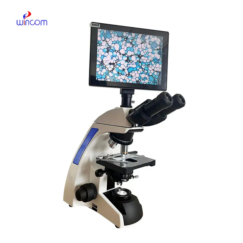
I’m looking to purchase several microscopes for a research lab. Please let me know the price list ...
Hello, I’m interested in your water bath for laboratory applications. Can you confirm the temperat...
E-mail: [email protected]
Tel: +86-731-84176622
+86-731-84136655
Address: Rm.1507,Xinsancheng Plaza. No.58, Renmin Road(E),Changsha,Hunan,China

 af
af
 es
es
 ar
ar
 tr
tr
 sw
sw
 pt
pt
 th
th
 ur
ur
 bn
bn
 ne
ne
 vi
vi
 km
km
 lo
lo
 de
de
 ru
ru
 fi
fi
 nl
nl
 fa
fa
 fr
fr
 ko
ko