Today, clinical laboratories always rely on hplc basics for the purpose of giving comprehensive chemical and biological data from patient samples. The technology's exceptional sensitivity and accuracy make it possible to separate even the smallest amounts of substances such as drugs and metabolites from complicated mixtures. Laboratory staff performs using hplc basics in method development, validation and ongoing monitoring of the lab's analytical performance. The multi-use of the instrument guarantees its presence during both normal testing and research work, hence hospitals and laboratories are always consistent in providing accurate and trustworthy diagnostic and analytical results.

The quality control process for hplc basics in intravenous medications and hospital-prepared solutions is being carried out by hospital laboratories. It isolates the impurities and analyzes the active substances to ascertain the uniformity of the composition. This practice enables the pharmacists and laboratory staff to verify the drug's quality before it gets to the patient, hence minimizing the risk associated with it and at the same time endorsing the safe therapeutic practices in hospitals.
Hospital laboratories will largely benefit from hplc basics systems that are meant for increased throughput and multi-sample analysis. The future instruments will merge improved sensitivity with strong automation, thus making rapid diagnostics and continuous monitoring of patient medications and metabolic profiles possible, which in turn will provide hospitals with safer and more efficient operations.

hplc basics will require regular maintenance to be kept up in order to continue providing precise measurements in medical laboratories. After every use, the technicians should flush the columns, check the seals, and inspect the tubing for wear and tear and ensure that the detector is working. Regular calibration and good solvent management decrease the chances of system damage and increase the consistency of the results. Good care and maintenance not only increase the efficiency of the laboratory but also help in providing reliable diagnostics and maintaining the instruments for hospital applications.
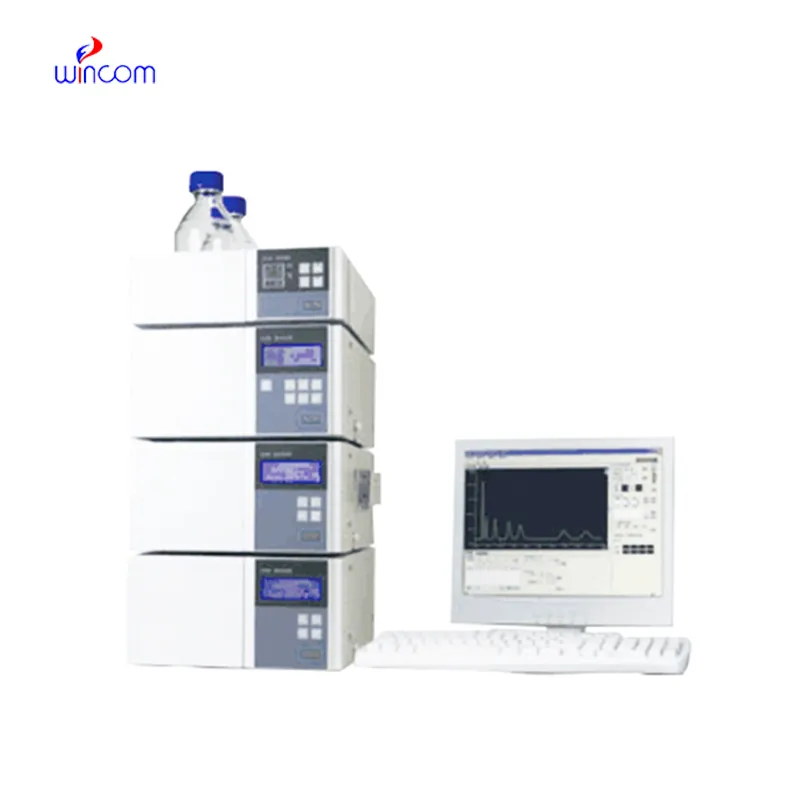
In today's laboratories, hplc basics is indispensable for chemical analysis and serves as a primary instrument. Detection of compounds in intricate mixtures is first done through separation and then identification. Consequently, researchers can precisely check the interactions between molecules. hplc basics is regarded to have extremely high reproducibility and it shares its strength with the fields of pharmaceuticals, biochemistry, and environmental science. Its alliance with sensitive detectors leads to the accurate measurement of very small amounts. hplc basics is the trustworthy partner of lab technicians in validation of experiments, profiling of samples, and development of analytical methods. It not only gives consistent and detailed results but also boosts the efficiency of laboratories and at the same time, makes sure that the data obtained from research is reliable and thus, supports the advanced scientific inquiries that are conducted in various disciplines.
Q: What is the sample preparation for HPLC? A: For the most part, samples should be filtered, diluted, or subjected to solvent extraction in order to avoid column clogs and have the results be accurate Q: Is HPLC able to pick trace-level compounds? A: With the right detectors, it can pick up such substances in extremely small amounts with high sensitivity. Q: Is HPLC a method that can be applied to analysis of proteins? A: Yes, particularly if one employs size-exclusion and reversed-phase columns for protein, peptide, and biomolecule separation. Q: What is the process of calibrating HPLC? A: The process is done by taking standards of known concentrations that are the same as the one in the sample and using them to check the performance of the column and the accuracy of the detector. Q: Are particular solvents needed for HPLC? A: Yes, the solvents used need to be compatible with the type of the column and the detectors to prevent any damage or interference in the analysis process.
The hospital bed is well-designed and very practical. Patients find it comfortable, and nurses appreciate how simple it is to operate.
The microscope delivers incredibly sharp images and precise focusing. It’s perfect for both professional lab work and educational use.
To protect the privacy of our buyers, only public service email domains like Gmail, Yahoo, and MSN will be displayed. Additionally, only a limited portion of the inquiry content will be shown.
Could you please provide more information about your microscope range? I’d like to know the magnif...
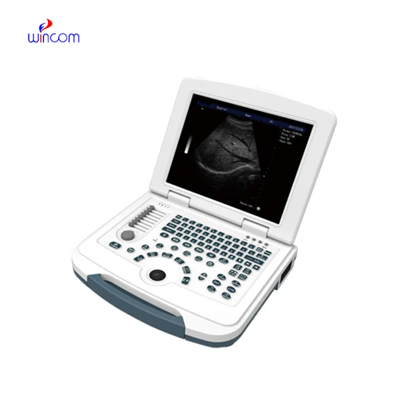
We’re currently sourcing an ultrasound scanner for hospital use. Please send product specification...
E-mail: [email protected]
Tel: +86-731-84176622
+86-731-84136655
Address: Rm.1507,Xinsancheng Plaza. No.58, Renmin Road(E),Changsha,Hunan,China

 af
af
 es
es
 ar
ar
 tr
tr
 sw
sw
 pt
pt
 th
th
 ur
ur
 bn
bn
 ne
ne
 vi
vi
 km
km
 lo
lo
 de
de
 ru
ru
 fi
fi
 nl
nl
 fa
fa
 fr
fr
 ko
ko