reversed phase hplc hangs the hospital laboratory in the sense of getting quick and reproducible results for patient sample analysis. Its use is widespread to separate small molecules, hormones, and therapeutic drugs with pinpoint accuracy. Lab staff apply reversed phase hplc in discovering biomarkers, doing pharmacokinetic studies, and metabolite profiling. Its flexibility makes it suitable for clinical applications with different requirements like research, routine diagnostics, and patient care. So, when hospitals include reversed phase hplc into their laboratory processes, they get not only the speed but also the dependable analytical performance over various departments.

Biochemical and clinical laboratories use reversed phase hplc to examine plasma or serum metabolites for disease research. It isolates and measures the amounts of small molecules participating in metabolism thus shedding light on patient conditions. The method is commonly employed in metabolic studies and experimental clinical trials conducted in hospitals.
Advanced software platforms for predictive analytics in healthcare are going to be part of the reversed phase hplc integration. The hospitals will take advantage of the real-time data provided by the patient samples to influence their clinical decisions. Molecular profiling as well as automated quality control and laboratory efficiency will be thereversed phase hplc future applications targeting the improvement of patient care.
The hospital labs keep their reversed phase hplc by adopting diligent handling and preventive maintenance. The regular examination of the columns, pumps, and connectors, along with the correct use of the solvents, aids in eliminating the problems of blockages and pressure. The lab staff is recommended to observe the cleaning and calibration according to the manufacturer's manual. The, such practices are applied, they bring about the benefits of long-term reliability, consistent separation quality, and accurate analytical outcomes in both clinical and experimental workflows.
Therapeutic drug monitoring relies heavily on reversed phase hplc in hospital settings. It determines the concentration of drugs in the body to guarantee efficiency and security. The laboratory staff uses it for the examination of blood, serum, or urine samples, and signifies small molecular compounds with high accuracy. By yielding consistent outcomes, reversed phase hplc services the medics in changing the amounts and preventing side effects. Its use goes to hormone level testing, metabolite analysis, and pharmacokinetics research. With quick processing and accurate information, reversed phase hplc is a part of the hospital patient care, making evidence-based treatment decisions possible and enhancing clinical outcomes in different departments.
Q: What types of HPLC columns are available? A: Reversed-phase, normal-phase, ion-exchange, and size-exclusion columns are the main types of columns used according to the nature of the analytes. Q: Can multiple samples be analyzed simultaneously? A: Yes, in high-throughput systems, automated sample injection and sequential analysis are among the techniques to achieve this. Q: How does temperature affect HPLC performance? A: Temperature changes can cause variations in separation efficiency and retention times; however, the majority of labs make use of precise temperature control. Q: Can HPLC be integrated with data software? A: Sure, it can be linked with laboratory software for data collection, processing, and reporting. Q: What types of laboratories use HPLC? A: HPLC is employed by hospitals, pharmaceuticals, biochemistry research, and environmental testing labs.
This x-ray machine is reliable and easy to operate. Our technicians appreciate how quickly it processes scans, saving valuable time during busy patient hours.
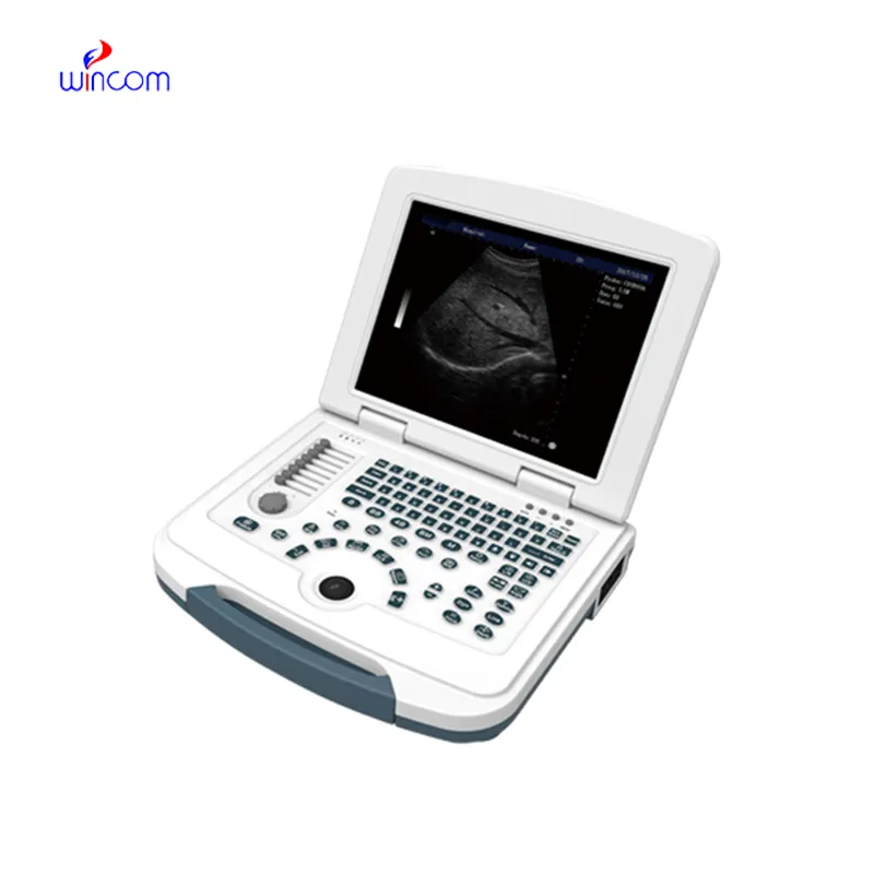
This ultrasound scanner has truly improved our workflow. The image resolution and portability make it a great addition to our clinic.
To protect the privacy of our buyers, only public service email domains like Gmail, Yahoo, and MSN will be displayed. Additionally, only a limited portion of the inquiry content will be shown.
Could you share the specifications and price for your hospital bed models? We’re looking for adjus...
I’d like to inquire about your x-ray machine models. Could you provide the technical datasheet, wa...
E-mail: [email protected]
Tel: +86-731-84176622
+86-731-84136655
Address: Rm.1507,Xinsancheng Plaza. No.58, Renmin Road(E),Changsha,Hunan,China

 af
af
 es
es
 ar
ar
 tr
tr
 sw
sw
 pt
pt
 th
th
 ur
ur
 bn
bn
 ne
ne
 vi
vi
 km
km
 lo
lo
 de
de
 ru
ru
 fi
fi
 nl
nl
 fa
fa
 fr
fr
 ko
ko