With multi-layer coated optics, the virus under microscope delivers better light transmission and image contrast. Ergonomic design allows for comfortable long-term use. The smooth stage movement and fine focusing system provide sensitive slide control for accurate analysis. The virus under microscope can be used with image capture systems for recording and sharing information, supporting both live observation and digital research workflows in the classroom and lab.

Applications of the virus under microscope cross into different spheres. It enables disease diagnosis by examining tissue sample and blood smears in medicine. In materials science, the virus under microscope is employed to examine crystal structures, coatings, and composites. In life sciences research, it is used in visualization of cell morphology, patterns of growth, and intracellular action. The virus under microscope also offers quality inspection for production with precision in semiconductor fabrication and microfabrication. It is used in museums and conservation laboratories to examine pigments and fibers in artifacts from ancient times.

With the progress of technology, the virus under microscope will turn into a smarter and more interactive research tool. Compatibility with AI will allow it to detect patterns, recognize anomalies, and measure data automatically. The virus under microscope will also make remote diagnostics possible, where the samples from every corner of the world can be diagnosed remotely by specialists. Advances in imaging sensors and optical systems will provide better depth resolution and faster capture rates. These will expand the uses of the virus under microscope in medicine, nanotechnology, and education.

In the interest of precision and reliability, the virus under microscope should be constantly exposed to cleanliness and maintenance. Switch it off at all times before adjusting or cleaning parts. The lenses may be cleaned with alcohol-free cleaners lightly to avoid scratching. Rotary components such as knobs and stage mechanisms value light lubrication at regular intervals. The virus under microscope must be stored away from direct sunlight and vibration. Professional checking once a year ensures optical alignment is not affected and prevents wear from invisible damage.
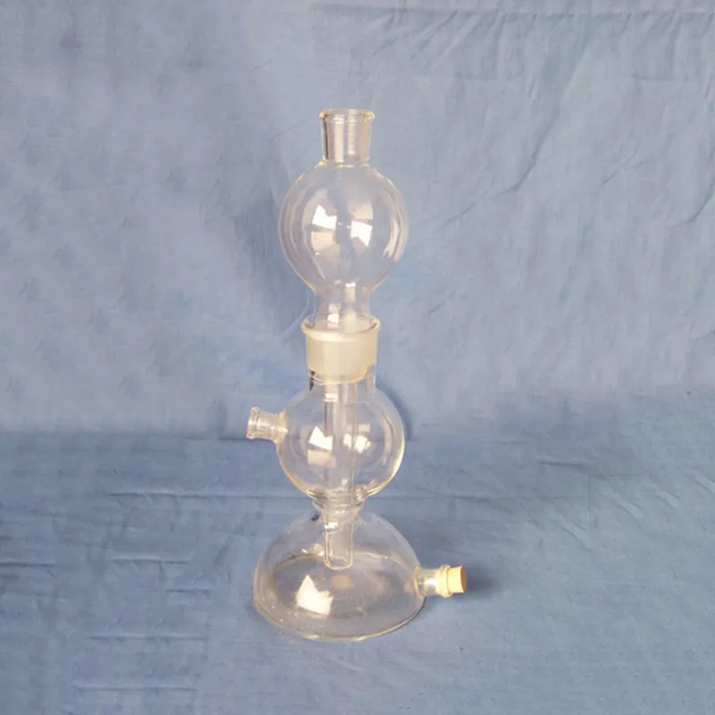
The virus under microscope bridges the visible and invisible by rendering small particles and organisms visible. Using a lens system and controlled light, the virus under microscope enables scientists and students to study samples with utmost precision. It has diverse applications in medicine, biology, electronics, and quality control. Digital and fluorescence forms extend study accuracy, simplifying visualization and data recording in most areas of science.
Q: What are the main parts of a microscope? A: The key components include the eyepiece, objective lenses, stage, focusing knobs, and illumination system, all working together to magnify and clarify specimens. Q: How do you clean the lenses of a microscope? A: Lenses should be cleaned using soft lens paper or microfiber cloth with a small amount of lens cleaner to avoid scratching or damaging optical coatings. Q: What magnification levels can a microscope achieve? A: Depending on the model, a microscope can typically achieve magnifications ranging from 40x to over 1000x for detailed observation of microscopic structures. Q: Why is light adjustment important in a microscope? A: Proper light adjustment ensures accurate contrast and brightness, allowing clear observation without distortion or glare during viewing. Q: Can a microscope be used for educational purposes? A: Yes, microscopes are widely used in classrooms and laboratories to teach students about biology, materials science, and microscopic analysis.
This x-ray machine is reliable and easy to operate. Our technicians appreciate how quickly it processes scans, saving valuable time during busy patient hours.
The centrifuge operates quietly and efficiently. It’s compact but surprisingly powerful, making it perfect for daily lab use.
To protect the privacy of our buyers, only public service email domains like Gmail, Yahoo, and MSN will be displayed. Additionally, only a limited portion of the inquiry content will be shown.
Could you share the specifications and price for your hospital bed models? We’re looking for adjus...
We’re interested in your delivery bed for our maternity department. Please send detailed specifica...
E-mail: [email protected]
Tel: +86-731-84176622
+86-731-84136655
Address: Rm.1507,Xinsancheng Plaza. No.58, Renmin Road(E),Changsha,Hunan,China

 af
af
 es
es
 ar
ar
 tr
tr
 sw
sw
 pt
pt
 th
th
 ur
ur
 bn
bn
 ne
ne
 vi
vi
 km
km
 lo
lo
 de
de
 ru
ru
 fi
fi
 nl
nl
 fa
fa
 fr
fr
 ko
ko