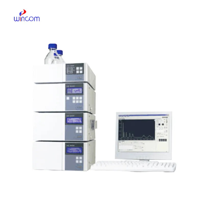
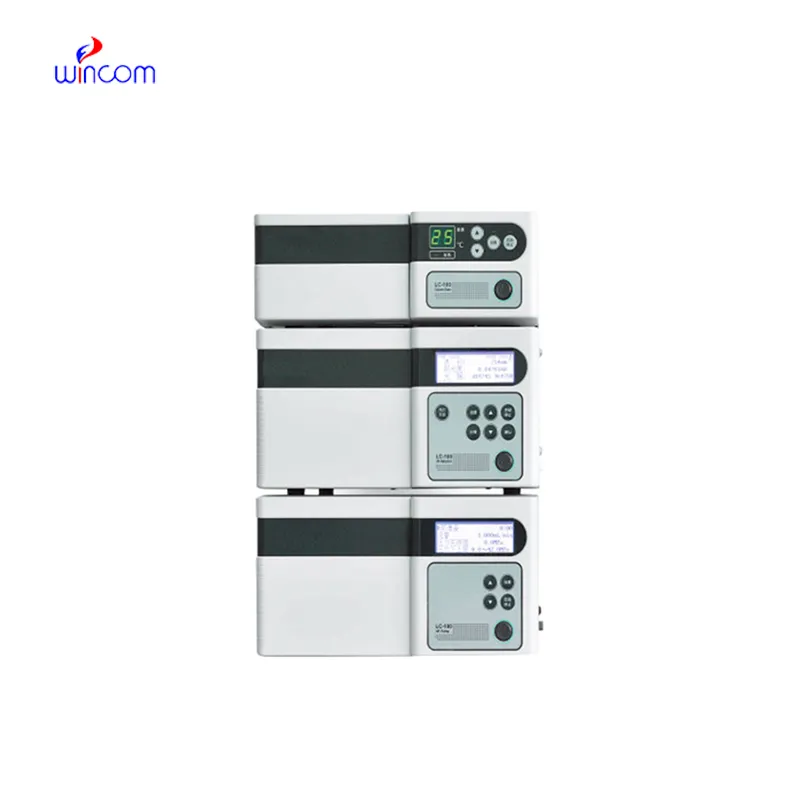
In the pharmaceutical lab, waters hplc system is the key to the precise assessment of the active substances, impurities, and metabolites. The machine gives a high-resolution separation, which in turn supports the quality assurance and the regulation compliance. Lab workers put their trust on waters hplc system for method validation, production consistency monitoring, and research trials. waters hplc system brings together the delicate ability to detect plus the repeated nature of results to make the complex formulations proficiently analyzed, thus, it serves the routine lab testing and the advanced experimental work in hospitals, research centers, and clinical facilities both.

Biochemical and clinical laboratories use waters hplc system to examine plasma or serum metabolites for disease research. It isolates and measures the amounts of small molecules participating in metabolism thus shedding light on patient conditions. The method is commonly employed in metabolic studies and experimental clinical trials conducted in hospitals.
Hospital laboratories will largely benefit from waters hplc system systems that are meant for increased throughput and multi-sample analysis. The future instruments will merge improved sensitivity with strong automation, thus making rapid diagnostics and continuous monitoring of patient medications and metabolic profiles possible, which in turn will provide hospitals with safer and more efficient operations.
The effectiveness of a laboratory is determined by the proper maintenance of waters hplc system. If the pump seals are regularly cleaned, the flow rates are monitored, and the usage of incompatible solvents is avoided then damage to the laboratory equipment can be prevented. It is essential for the technicians to carefully examine the columns, detectors, and tubing and in case of any sign of wear to conduct the scheduled calibration. Keeping waters hplc system in their best condition guarantees reproducibility, lowers the risk of equipment breakdown, and provides continuous performance for both hospital tests and experiments.
In today's laboratories, waters hplc system is indispensable for chemical analysis and serves as a primary instrument. Detection of compounds in intricate mixtures is first done through separation and then identification. Consequently, researchers can precisely check the interactions between molecules. waters hplc system is regarded to have extremely high reproducibility and it shares its strength with the fields of pharmaceuticals, biochemistry, and environmental science. Its alliance with sensitive detectors leads to the accurate measurement of very small amounts. waters hplc system is the trustworthy partner of lab technicians in validation of experiments, profiling of samples, and development of analytical methods. It not only gives consistent and detailed results but also boosts the efficiency of laboratories and at the same time, makes sure that the data obtained from research is reliable and thus, supports the advanced scientific inquiries that are conducted in various disciplines.
Q: What is the sample preparation for HPLC? A: For the most part, samples should be filtered, diluted, or subjected to solvent extraction in order to avoid column clogs and have the results be accurate Q: Is HPLC able to pick trace-level compounds? A: With the right detectors, it can pick up such substances in extremely small amounts with high sensitivity. Q: Is HPLC a method that can be applied to analysis of proteins? A: Yes, particularly if one employs size-exclusion and reversed-phase columns for protein, peptide, and biomolecule separation. Q: What is the process of calibrating HPLC? A: The process is done by taking standards of known concentrations that are the same as the one in the sample and using them to check the performance of the column and the accuracy of the detector. Q: Are particular solvents needed for HPLC? A: Yes, the solvents used need to be compatible with the type of the column and the detectors to prevent any damage or interference in the analysis process.
The centrifuge operates quietly and efficiently. It’s compact but surprisingly powerful, making it perfect for daily lab use.
This x-ray machine is reliable and easy to operate. Our technicians appreciate how quickly it processes scans, saving valuable time during busy patient hours.
To protect the privacy of our buyers, only public service email domains like Gmail, Yahoo, and MSN will be displayed. Additionally, only a limited portion of the inquiry content will be shown.
I’m looking to purchase several microscopes for a research lab. Please let me know the price list ...
Hello, I’m interested in your centrifuge models for laboratory use. Could you please send me more ...
E-mail: [email protected]
Tel: +86-731-84176622
+86-731-84136655
Address: Rm.1507,Xinsancheng Plaza. No.58, Renmin Road(E),Changsha,Hunan,China

 af
af
 es
es
 ar
ar
 tr
tr
 sw
sw
 pt
pt
 th
th
 ur
ur
 bn
bn
 ne
ne
 vi
vi
 km
km
 lo
lo
 de
de
 ru
ru
 fi
fi
 nl
nl
 fa
fa
 fr
fr
 ko
ko