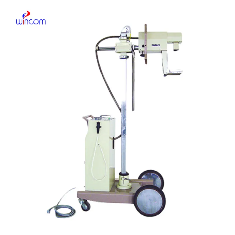
The welding x ray machine integrates the principles of ergonomics with high-quality imaging technologies. The easy-to-use system enables users to navigate with ease. The system also integrates automatic calibration functionality to ensure accurate outcomes. The welding x ray machine supports digital storage and retrieval solutions that offer healthcare providers easy access to diagnostic images.

The welding x ray machine is used extensively in dentistry, yielding minute-level images of teeth, jaw bones, and surrounding tissues. It assists in the diagnosis of cavities, orthodontic problems, and impacted tooth diagnosis. The welding x ray machine is used for endodontic and implant planning to allow precision treatment delivery.
In the years to come, the welding x ray machine shall be at the forefront of predictive and personalized medicine. Smart imaging platforms shall look into patient history in conjunction with real-time scans in an attempt to predict upcoming health problems. The welding x ray machine shall therefore turn into an anticipatory diagnostic system instead of a reactive one.

Care and maintenance of the welding x ray machine are required to ensure repeat imaging quality and ruggedness. Cable, detector, and collimator faults are averted by periodic checks. The welding x ray machine need to be kept in a dust-free environment with low temperatures to avoid overheating and dust depositing on them. Routine calibration and radiation output monitor checks ensure accurate diagnostic data.
The welding x ray machine uses X-ray transmission through the body to form an image on a detector that helps the doctor see the inside of the body without resorting to surgical procedures. The welding x ray machine produces images that have high clarity and resolution to ensure accurate diagnoses. The welding x ray machine has various applications in medicine depending on the part of the body that needs to be viewed.
Q: What makes an x-ray machine different from a CT scanner? A: An x-ray machine captures a single 2D image, while a CT scanner takes multiple x-rays from different angles to create 3D cross-sectional views. Q: How is image quality measured in an x-ray machine? A: Image quality depends on factors like contrast, resolution, and exposure settings, which are adjusted based on the target area being examined. Q: What power supply does an x-ray machine require? A: Most x-ray machines operate on high-voltage power systems, typically between 40 to 150 kilovolts, depending on their intended use. Q: Can x-ray machines be used for dental imaging? A: Yes, specialized dental x-ray machines provide detailed images of teeth, jaws, and surrounding structures to support oral health assessments. Q: How does digital imaging improve x-ray efficiency? A: Digital systems allow instant image preview, faster diagnosis, and reduced need for retakes, improving workflow efficiency in clinical environments.
The microscope delivers incredibly sharp images and precise focusing. It’s perfect for both professional lab work and educational use.
We’ve been using this mri machine for several months, and the image clarity is excellent. It’s reliable and easy for our team to operate.
To protect the privacy of our buyers, only public service email domains like Gmail, Yahoo, and MSN will be displayed. Additionally, only a limited portion of the inquiry content will be shown.
We’re looking for a reliable centrifuge for clinical testing. Can you share the technical specific...
Hello, I’m interested in your centrifuge models for laboratory use. Could you please send me more ...
E-mail: [email protected]
Tel: +86-731-84176622
+86-731-84136655
Address: Rm.1507,Xinsancheng Plaza. No.58, Renmin Road(E),Changsha,Hunan,China

 af
af
 es
es
 ar
ar
 tr
tr
 sw
sw
 pt
pt
 th
th
 ur
ur
 bn
bn
 ne
ne
 vi
vi
 km
km
 lo
lo
 de
de
 ru
ru
 fi
fi
 nl
nl
 fa
fa
 fr
fr
 ko
ko